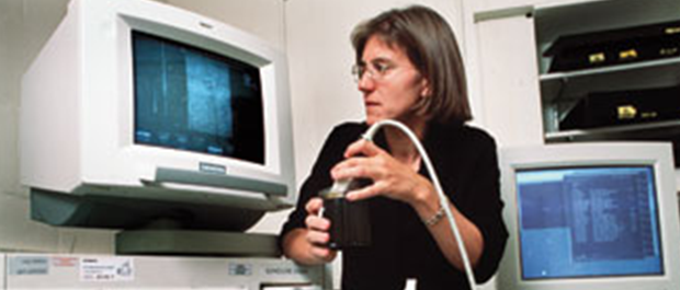
 Mysteries of the heart: Medical diagrams, such as this one from 1875, could help diagnosticians only so much; more than a century later, it’s gotten easier, thanks to ultrasound. Corbis The elderly man lies on the examining table, his expression taut with pain and fear. Earlier that morning, a crushing angina attack left him clutching his chest in panic. Placing a small ultrasound wand onto the man’s chest, the cardiologist peers up at a wall-sized screen in the examining room. A three-dimensional image materializes, showing the man’s pulsing coronary arteries, like a flowing river system with its winding tributaries. The physician touches a joystick to zoom in on the arterial landscape, pinpointing the blockages and providing a map for surgery that will save the man’s heart and his life. In a remote South American forest, a white van lurches along the rutted dirt road into a village and comes to a stop. A group of women greets the mobile screening clinic, filing into the van for their examinations. They take turns on the examining table as the clinic nurse guides an ultrasound wand over each woman’s breasts. The nurse scrutinizes a computer screen showing a 3D image of each breast. Several of the women have lumps in their breasts; the ultrasound analysis reveals most to be nothing more than benign, fluid-filled cysts. But the lump in one woman’s breast shows tell-tale characteristics of a solid tumor. The nurse types commands into a laptop computer. A small robotic arm equipped with a biopsy needle glides smoothly into position. The robot pin-points the suspicious mass via an ultrasound array in its tip. Guided by artificial intelligence, the robot eases the biopsy needle gently and precisely into the breast to extract a tissue sample. Soon the woman will know whether her worries are justified. These scenarios represent dreams of Duke bioengineers who, over four decades, have invented technology that transformed ultrasound imaging from a laboratory curiosity into an essential clinical instrument. They’re dreams for now, but today, clinicians routinely use ultrasound to provide images of fetuses, as well as the heart and other organs. Over the coming decades, researchers expect ultrasound to influence medicine even more profoundly— including diagnosing and treating heart disease, liver disease, stroke, brain cancer, breast cancer, prostate cancer, and even battlefield wounds. Ultrasound imaging appeals to physicians because its basic technology is simple and benign. Ultrasound machines, unlike multi-ton MRI machines or room-sized CAT scanners, are typically no larger than a baby buggy and just as portable. And unlike X-rays or radioactive-tracer PET scans, ultrasound does not expose patients to ionizing radiation. Ultrasound is also far cheaper. Even the most elaborate ultrasound scanner costs no more than $100,000, versus millions of dollars for MRI or CAT scanners. A tour of the Duke biomedical engineering department’s ultrasound labs— where some of the field’s most important technology was born—reveals a collection of equipment as exotic and, yes, sometimes peculiar as any creative research laboratory. Visitors may expect to see electronics-assembling clean rooms, robotic arms, and circuit-slicing saws with hair-thin diamond blades that spin at 30,000 rpm. But they might not expect to see turkey breasts or a garbage can full of murky river water—both of which have figured in key Duke experiments. Ultrasound—sound at a frequency higher than the human ear can detect— had been in scientific use largely as a laboratory tool for studying the body since the 1940s. However, the creative ingenuity that transformed the technology into everyday clinical instruments began at Duke in 1967. That year, Theo Pilkington M.S. ’60, Ph.D. ’63, a brash, young associate professor and the driving force behind biomedical engineering at Duke, recruited ultrasound engineer Frederick “Fritz” Thurstone to the new department. Thurstone was an avid proponent of medical ultrasound, although his earliest experiments were primitive. He would immerse patients in water and “ping” them with a parabolic reflector, much as submarines pinged enemy warships. But it was two of Thurstone’s early graduate students—Olaf von Ramm Ph.D. ’73 and Stephen Smith Ph.D. ’75—who would join with him to invent technology that would propel ultrasound into just about every medical center in the world.  Advanced at the time: Early ultrasound testing using Thurstone’s sixteen-transducer prototype. Courtesy Olaf von Ramm One major aim was to revolutionize a device called the transducer, which serves as both the transmitter and receiver of ultrasound pulses. Thurstone and von Ramm advanced transducer technology: What had been simple devices that emitted a single pulse became multiple arrays that produced up to thousands of pulses at once. The intricate reflections of these pulses from body tissues and organs could be processed by computers into the first ultrasound images. In their work, the engineers collaborated closely with Duke physicians. When the cardiologists saw the first ultrasound image of a beating heart, “they didn’t know what they were looking at,” von Ramm recalls. “Their mental image was based on static X-rays. So, we all learned together what a living heart looked like.” Ultrasound enabled cardiologists to measure noninvasively a heart’s “ejection fraction”—the volume of blood that it pumps. No catheter insertion was needed, and no X-rays required, to obtain this critical measure of heart function.  Internal perspective: Scan of a heart from the original ultrasound machine at Duke. Courtesy Olaf von RammThe next major advance in ultrasound technology was triggered by a plane crash. In 1982, an Air Florida jet slammed into the Potomac River, sinking immediately. Divers could not find bodies in the dark waters. Hearing of the tragedy, Smith envisioned that a 3D ultrasound machine would allow search parties to “see” in real time what was at the bottom of murky bodies of water. Smith and von Ramm set out to invent such a machine, developing complex transducer arrays and fast computer systems that could analyze the cascade of ultrasound reflections and produce a three-dimensional moving ultrasound image in real time. In 1987, they realized their vision. In early experiments, they carted a garbage can full of brownish water from the nearby Eno River back to the lab and used a submerged wrench as their target. They could locate the wrench using the new technology. With the success of their system, they joined with partner John Oxaal B.S.E. ’76 to form Volumetrics Medical Imaging to develop the first real-time 3D commercial system. Since then, 3D ultrasound technology has come into wide clinical use for imaging tissues and organs. But ultrasound technology has the potential to produce an even more detailed view of the body. Von Ramm’s vision of the future is to use advanced imaging processing and high-frequency transducers to create high-resolution images of even smaller segments of tissue, down to the level of the coronary arteries. “I want to see the coronary artery tree hanging in 3D space,” he says. “We’ve imaged them for short distances, but they are very tortuous vessels that curve around the heart, and the heart is a 3D object.” Smith and staff engineer Ned Light B.S.E. ’89, M.S.E. ’97 have continued to develop more specialized ultrasound probes, concentrating particularly on smaller ones that can be inserted into the body for a clearer view of internal organs. They first invented endoscopic probes that could fit into the throat or through incisions in the body. But their latest project has gotten even smaller. In his laboratory, Light holds up a thin tube—a catheter that surgeons thread into the body to carry such devices as heart valves and tiny clot-blocking wire cages to be inserted into blood vessels. But the tip of this catheter is different from ordinary devices. It has embedded in it a ring of tiny wires. These wires are ultrasonic transducers that will act like the equivalent of a flashlight beaming pulses. This “flashlight” will give surgeons a close-up view of the progress of the catheter through blood vessels, as well as the procedure they are conducting. Such devices could reduce or eliminate the need for Xray imaging and potentially toxic dyes currently used to guide such procedures. Like all creative researchers, Duke ultrasound engineers think in “what-ifs,” then set out to make them reality. Among them: What if oncologists could insert an ultrasound probe into a brain tumor, not only to image the tumor but to kill it as well? Graduate student Carl Herickhoff M.S.E. ’09, Ph.D. ’11, a member of Smith’s lab, is experimenting with an ultrasound-equipped catheter that could be inserted into the brain to locate a tumor. Physicians hypothesize that they might then apply more power to the ultrasound probe to gently heat the tumor. The patient would already have been injected with cancer-killing chemicals encapsulated in heat-sensitive fatty bubbles called liposomes. The ultrasound heating would be just enough to melt the bubbles in the tumor, delivering a targeted chemotherapy dose. What if stroke victims could be diagnosed in the home or ambulance and clot-busting therapy given immediately? Such quick treatment would more likely fall within the “golden hours,” during which vital brain functions can be saved. To enable such rapid treatment, Smith and graduate student Brooks Lindsey are developing a 3D ultrasound “brain helmet” that emergency technicians would fit onto a stroke victim’s head. The helmet could transmit an ultrasound brain image to the hospital neurologist and immediately show whether the stroke was caused by a clot or a bleeding vessel. If it were a clot, the doctor could prescribe clotbusting drugs to be given on the way to the hospital, rather than waiting to perform a CT scan. What if ultrasound could distinguish potential breast cancers from harmless cysts? And what if that imaging could guide a robotic arm to obtain a biopsy sample? Using an ultrasound wand fitted on a commercially available robot arm, bioengineering student Kaicheng Liang B.S.E.’10, working in Smith’s lab, has shown that the device can locate an artificial cyst in flesh—in this case, a turkey breast. The next step is to test how well the ultrasound system can locate real breast tumors, compared to physicians relying on 2D ultrasound or mammograms. Liang and his colleagues are also exploring whether such a robotic system could locate and take samples of prostate tissue. What if ultrasound could guide a robotic arm to extract shrapnel on the battlefield? Albert J. Rogers B.S.E. ’09 has shown that an alternating magnetic field will subtly vibrate tiny bits of metal in water, rendering them visible enough on ultrasound to guide the tip of a robotic arm to their location. The finding raises the promise of an artificially intelligent battlefield surgeon that could deftly remove shrapnel quickly before it does more damage. What if ultrasound could not only “see” tissues but also “feel” them? Physicians’ sense of touch is one of their most valuable diagnostic tools, as they palpate their patients’ bodies to detect tumors and other abnormalities. Bioengineering professors Gregg Trahey Ph.D. ’85 and Kathryn Nightingale B.S.E. ’89, Ph.D. ’97 and their colleagues are developing a technology called Acoustic Radiation Force Impulse (ARFI) imaging, which uses strong ultrasound pulses to “poke” tissues or organs deep in the body. The tissue movement is infinitesimal— about a hundredth of the diameter of a human hair. But the ultrasensitive ultrasonic detection system is designed to detect the subtle reflected signals from these tiny motions—amidst the relatively huge motions of the heart and other organs—to measure the stiffness of the target tissue.  Exquisite specificity: Kathy Nightingale, above left, uses ultrasound imaging for detailed diagnostics; ultrasound image of a canine brain, above, shows blood vessels in color and images of lateral ventricles (LV), base of the skull (B), internal carotid artery (ICA), and anterior cerebral artery (ACA). Top: University Photography; Bottom: Courtesy Stephen Smith Trahey recalls that a phone call from the director of Duke’s breast cancer clinic in 1993 triggered him and his colleagues to consider ARFI as a clinical tool. Using ultrasound, the clinic’s director complained, “We can’t even do the simplest thing. We can’t tell the difference between a cyst and a solid lesion. You guys are worthless.” Nightingale, a graduate student of Trahey’s, set out to explore whether ARFI’s poking mechanism could distinguish between harmless fluid-filled cysts and potentially malignant solid masses. She found that the technique worked, and she is now exploring whether ARFI can more definitively spot malignant tumors. “Some cancers and some benign masses can be similar in stiffness,” she says. “But cancers tend to be more tethered within the breast and prostate, more connected to the surrounding tissue. So, we have begun exploring techniques to highlight the differences in order to distinguish cancers.” ARFI’s ultrasonic poking can also tell squishy living tissue from stiffer dead tissue. Trahey, fellow bioengineer Patrick Wolf Ph.D. ’92, and cardiologist Tristram Bahnson are developing ARFI probes that could guide cardiologists when they perform the tissue-killing procedure called cardiac ablation. In this procedure, they use a probe that emits intense radio frequency waves to kill bits of heart tissue selectively and to block the dangerous wave of arrhythmia moving through the heart. “Currently they fly blind,” Trahey says. “You can’t really tell which part of the heart you’ve cooked and which part is intact. They watch the patient’s arrhythmia and use invasive electroanatomic mapping techniques, and some of these procedures can take eight, ten, twelve hours.” Nightingale is working with Duke hepatologists to explore ARFI as a technique to detect fibrosis in the liver, a sign of cirrhosis and elevated risk of liver cancer. “Given that the technique is noninvasive, it could be preferable to needle biopsies, in which a plug of tissue is extracted and the overall extent of fibrosis inferred,” she says. “The needle might land in a ‘bad neighborhood’ that doesn’t accurately portray the overall status of the liver, while the ARFI method can be performed painlessly, in multiple locations.” For Nightingale, a related interest is whether ARFI could guide biopsies of the prostate to detect cancer. The technique would be a high-tech version of the physician’s digital rectal exam, she says. “What physicians do in those exams is to feel for regions that are stiffer, but biopsies are systematically taken from a grid of locations in the prostate. So, the idea is that we could use ARFI to identify regions where they should aim their biopsy needle." Anaesthesiologists could use ARFI to guide a needle administering regional anesthesia. Nightingale and assistant research professor Mark Palmeri B.S.E. ’00, Ph.D. ’05, M.D. ’07 are developing ARFI techniques to map the change in stiffness of tissue as the needle infuses anaesthetic into a region. Such guidance would enable anaesthesiologists to more precisely place a needle to deaden nerves more effectively. Faster processing and improved transducers are enabling von Ramm and his colleagues to track ever-finer motions of the heart. Such advances, they believe, will reveal— at cell-level detail—the instantaneous wave of contraction sweeping across the heart. Seeing the finest details of a heartbeat means they could pinpoint the regions of the heart damaged by heart attacks or the effects of congestive heart failure. Trahey and his colleagues are experimenting with a new imaging strategy that promises to greatly enhance clarity of ultrasound scans—just as high-def TV has superseded low-def. Called “coherence imaging,” the technique involves forming images not just from the ultrasound reflections themselves, but also from the reflections of finer “wavelets” formed when one ultrasound wave interferes with another. Trahey, research professor Jeremy Dahl Ph.D. ’04, and graduate student Muyinatu Lediju are developing the concept. In the view of George Truskey, chair of the biomedical engineering department, ultrasound and other technologies can be a big factor in a changing health-care landscape— notably by reducing the number of expensive and invasive biopsies. “A lot of people do have the perception that technology drives up health-care costs, and there are certainly cases where technology is overused,” he says. The use of expensive MRI scans on patients who don’t really need them is an obvious example. “There will always be the caveat that you have to monitor how these technologies are used. But the flip side is that the technology benefits patients by providing early diagnosis. That can lead to successful treatment, to prolonging someone’s life, and to stretching out that individual’s productive years.”
Meredith is a science writer and research communication consultant. He is the author of Explaining Research: How to Reach Key Audiences to Advance Your Work (Oxford University Press, 2010). For the latest news from the B.M.E. department: http://bme.duke.edu/research/biomedical-imaging. |
Share your comments
Have an account?
Sign in to commentNo Account?
Email the editor